

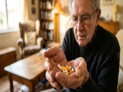
دونت هذه الملاحظةاسفل نتيجةفحص البول الكحول تاثيره اقوى على الخلاياالسليمةام الخبيثةففحوصات خلويةالبول لمرتين اصابتي ب(TCC) The simple will70 alcohol if might affect the cells better to be repeated with out Alcohol should arrive to the lab
هل ترغب في التحدث إلى طبيب نصياً آو هاتفياً؟
يمكنك الحصول على استشارة مجانية لأول مرة عند الاشتراك
2019-10-15 14:59:33
ممكن اعرف نتيجة التحليل دا ايه بالظبط يا دكتور sperm count in susceptible to measurement due to incomplete liquefaction of the semen sample oilgozoospermia
ممكن نتيجة التحليل دا ايه بالظبط يا sperm count in susceptible to measurement due to incomplete liquefaction of the semen sample oilgozoospermia
1
2017-12-17 16:35:36
ارجو التفسير REPORT Normal marrow signal of the lumbar vertebrae, with no focal lesions identified. Mild scoliosis of the lumbar spine with convexity to the left side D
التفسير REPORT Normal marrow signal of the lumbar vertebrae with no focal lesions identified Mild scoliosis of the lumbar spine with convexity to the left side D
1
2017-10-19 21:07:40
عملت اشعةصوتيةللخصيةواطلعت على النتائج فتبين لل اني سليم ماعدا هذه الجملة Both epididymides are unremarkable apart from simple cyst seen in the left epididymis measuring 10mm
عملت اشعةصوتيةللخصيةواطلعت على النتائج فتبين لل اني سليم ماعدا هذه الجملة Both epididymides are unremarkable apart from simple cyst seen in the left epididymis measuring mm
1
2013-11-11 18:40:13
ارجوكم ردوا اعاني من انعدام الحيوانات المنويه وهذا تقرير الاشعه عبر الشرج the prostate : there is about 1.5×1.3cm anechoic cystic lesion with echogenic irregular wall seen in the med portion of the prostate 12cm3 in volume with homogenous echogencity and no significant effect upon the base of the urinary bladder. VAS DEFERANCE both vas deferance come horizontally , join the ejaculatory ducts and end within the prostate urethra . their distal end is obscured by the previoussly mentioned lesion SEMINAL VESICLES both seminal vesieles measured about 5cm in long and 1.8cm in Ap diameter with echogenic content mostly due to the proximal obstruction by the previous mentioned lesion IMPRESSION: suspected a small abscess seen the central portion of the prostate as described above
كم ردوا اعاني من انعدام الحيوانات المنويه و تقرير الاشعه عبر الشرج the prostate there is about ×cm anechoic cystic lesion with echogenic irregular wall seen in the med portion of the prostate cm in volume with homogenous echogencity and no significant effect upon the base of the urinary bladder VAS DEFERANCE both vas deferance come horizontally join the ejaculatory ducts and end within the prostate urethra their distal end is obscured by the previoussly mentioned lesion SEMINAL VESICLES both seminal vesieles measured about cm in long and cm in Ap diameter with echogenic content mostly due to the proximal obstruction by the previous mentioned lesion IMPRESSION suspected a small abscess seen the central portion of the prostate as described above
2
يشير تقرير صورة البروستات إلى وجود خراج او دمل في جزء البروستات المركزي، وهذا الخراج يعمل على الضغط على الحويصلات المنويه لذلك لا تخرج الحيوانات المنوية الى الخارج فيكون السائل المنوي خالي من الحيوانات المنوية أو انعدام الحيوانات المنوية. لذلك ينصح بالذهاب إلى اخصائي مسالك بولية لعلاج خراج البروستات، حيث يتم العلاج من خلال المضاد الحيوي المناسب وتفريع الخراج الموجود في البروستات.
ومن أعراض خراج البروستات الذي يحدث بعد وجود التهاب بكتيري في البروستات ما يلي:
لذلك يجب عدم الانتظار ومراجعة الطبيب المختص خلال اقرب وقت للعلاج.
للمزيد:
المرجع:
2013-11-08 15:33:49
اعاني من انعدام الحيوانات المنويه بعد اجراء كل الفحوص لمعرفة السبب كانت النتيجه the prostate : there is about 1.5×1.3cm anechoic cystic lesion with echogenic irregular wall seen in the med portion of the prostate 12cm3 in volume with homogenous echogencity and no significant effect upon the base of the urinary bladder. VAS DEFERANCE both vas deferance come horizontally , join the ejaculatory ducts and end within the prostate urethra . their distal end is obscured by the previoussly mentioned lesion SEMINAL VESICLES both seminal vesieles measured about 5cm in long and 1.8cm in Ap diameter with echogenic content mostly due to the proximal obstruction by the previous mentioned lesion IMPRESSION: suspected a small abscess seen the central portion of the prostate as described above الحل؟
اعاني من انعدام الحيوانات المنويه بعد اجراء كل الفحوص لمعرفة السبب كانت النتيجه the prostate there is about ×cm anechoic cystic lesion with echogenic irregular wall seen in the med portion of the prostate cm in volume with homogenous echogencity and no significant effect upon the base of the urinary bladder VAS DEFERANCE both vas deferance come horizontally join the ejaculatory ducts and end within the prostate urethra their distal end is obscured by the previoussly mentioned lesion SEMINAL VESICLES both seminal vesieles measured about cm in long and cm in Ap diameter with echogenic content mostly due to the proximal obstruction by the previous mentioned lesion IMPRESSION suspected a small abscess seen the central portion of the prostate as described above الحل
1
2013-10-30 12:42:06
السلام عليكم ارجوكم اجيبوني ع هذا من عند الطبيب هل يوجد حصى ف الكليه او يوجد التهاب البروستاتا حاد هذي معلومات : procedure :K .U .B and prostate US. usg kub and prostate kidneys : both kidneys are normal in size, shape, axis and position. cortico-medullary ecotexture and thickness is normal. no focal lesion seen. mild fullness seen in both kidneys, no definite stone echo noted. urinary bladder : the bladder is well distended. no calcull is seen. the wall thickness is normal. the prostate is enlarged in size ( volume is 21.8 cc ) with hypoechoic echotexture, looks to be prostatitis impression: mild fullness in both kidneys prostatitis normal urinary bladder for clinical and lab corelation.. ارجوكم اجيبوني ع هذي المعلومات.. وشكرا
كم اجيبوني ع من عند الطبيب هل يوجد حصى ف الكليه او يوجد التهاب البروستاتا حاد هذي معلومات procedure K U B and prostate US usg kub and prostate kidneys both kidneys are normal in size shape axis and position cortico medullary ecotexture and thickness is normal no focal lesion seen mild fullness seen in both kidneys no definite stone echo noted urinary bladder the bladder is well distended no calcull is seen the wall thickness is normal the prostate is enlarged in size volume is cc with hypoechoic echotexture looks to be prostatitis impression mild fullness in both kidneys prostatitis normal urinary bladder for clinical and lab corelation كم اجيبوني ع هذي و
1
2013-10-22 22:06:50
هل هناك خطوره examination reveals clusters of small follicular , mixed with fibroblasts , and lymphocytes , the cells are monomorphic , with no cytological , no malignant cells could be detected follicular neoplasm no cytological malignaanc
هل خطوره examination reveals clusters of small follicular mixed with fibroblasts and lymphocytes the cells are monomorphic with no cytological no malignant cells could be detected follicular neoplasm no cytological malignaanc
1
2013-08-16 10:45:12
is there any way or sign that a mother could do or notice to know if her fetus is growning normally except the ultrasound?
is there any way or sign that a mother could do or notice to know if her fetus is growning normally except the ultrasound
3
2011-12-23 01:16:28
two short straight :one seen at the junction between the membranous and bulbar urethra with significant peri –stenotic dilatation of the membranous urethra. The other one seen at the region of the junction between the bulbar and penile urethra with also significant peri –stenotic dilatation. Evidence of bilateral vesico- urteric reflux changes and bilateral urteric and calyceal opacification. Normai appearance of the rest of penil urethra. اريد معرفة مضمون تقرير هذه الاشعة
two short straight one seen at the junction between the membranous and bulbar urethra with significant peri –stenotic dilatation of the membranous urethra The other one seen at the region of the junction between the bulbar and penile urethra with also significant peri –stenotic dilatation Evidence of bilateral vesico urteric reflux changes and bilateral urteric and calyceal opacification Normai appearance of the rest of penil urethra اريد معرفة مضمون تقرير هذه الاشعة
2
2010-06-11 09:12:28
12 اشعه دوبلر على الخصيتين لقد قمت بعمل اشعه دوبلر على الخصيتين نظرا لوجود دوالى والام شديده بالخصيتين ارجو منكم شرح تقريرالاشعه وما اذا كان يوجد ضمور بالخصيه وهل لا بد من عمل جراحه للدوالى بالخصيتين ام لا وهذ هو التقرير report gray-scal and color flow imaging of the scrotum revealed; gray-scale Left testis and epididymis; are of average size and homogenous texture with no focal lesions could be noted. right testis and epididymis; are of average size and homogenous texture with no focal lesions could be noted left pampinform venous plexus;presenting moderate dilatation of theveins where the maximamum diametre of the cross section of themost dilated vein posterior to the left testis measures about 3.1 mm during rest and 3.4mm at straining. left pampinform venous plexus;presenting normal caliber of he veins where the maximum diametre of the coss section of most prominent vein posterior to te right testis measures about 1.9mm during rest and 1.9mm at straining. .no hydrocele. color flow and pectral analysis .left pampinform venos plexus;exhibit posiive reflux on straining where the duration of the reflux measuring about 2sec. .right pampinform venos plexus;exhibt no reflux on straining. testicular arteries;appears patent with normal color flow and wavefom. opinion; .grade ll refluxing left varicocele normal both tests and epididymis
اشعه دوبلر على الخصيتين لقد قمت بعمل اشعه دوبلر على الخصيتين نظرا لوجود دوالى والام شديده بالخصيتين منكم شرح تقريرالاشعه وما اذا كان يوجد ضمور بالخصيه وهل لا بد من عمل جراحه للدوالى بالخصيتين ام لا وهذ هو التقرير report gray scal and color flow imaging of the scrotum revealed gray scale Left testis and epididymis are of average size and homogenous texture with no focal lesions could be noted right testis and epididymis are of average size and homogenous texture with no focal lesions could be noted left pampinform venous plexus presenting moderate dilatation of theveins where the maximamum diametre of the cross section of themost dilated vein posterior to the left testis measures about mm during rest and mm at straining left pampinform venous plexus presenting normal caliber of he veins where the maximum diametre of the coss section of most prominent vein posterior to te right testis measures about mm during rest and mm at straining no hydrocele color flow and pectral analysis left pampinform venos plexus exhibit posiive reflux on straining where the duration of the reflux measuring about sec right pampinform venos plexus exhibt no reflux on straining testicular arteries appears patent with normal color flow and wavefom opinion grade ll refluxing left varicocele normal both tests and epididymis
1
هل ترغب في التحدث مع طبيب؟
احصل الآن على استشارة مجانية لأول مرة عند الاشتراك في الخدمة
هل وجدت هذا المحتوى الطبي مفيداً؟
تحدث مع طبيب بسرية تامة و ناقش حالتك الصحية
مجاناً - احصل على إجابة لاستشاراتك الطبية
نخبة من الأطباء المتخصصين سيقومون بالإجابة على استفساراتك خلال 48 ساعة
5000 طبيب يستقبلون حجوزات عن طريق الطبي.
ابحث عن طبيب واحجز موعد في العيادة أو عبر مكالمة فيديو بكل سهولة
السماح للطبي بارسال التنبيهات والاشعارات التي تحتوي المعلومات الطبية والاجراءات الصحية من المحتوى الطبي
هل تريد التحدث مع طبيب الآن
اشترك الان واحصل على فتره مجانية
اشترك الان